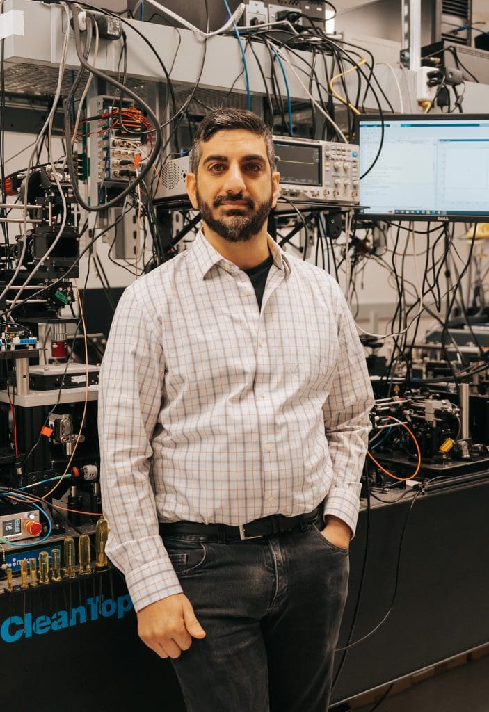
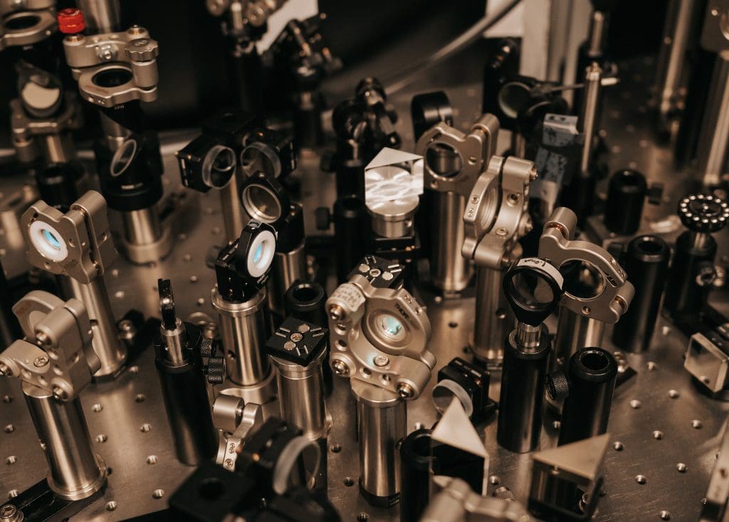
Spectroscopy is a broad field that studies the interaction of electromagnetic radiation and matter and is used across numerous disciplines, including chemistry, physics, biology, and astronomy.
 “We use electromagnetic radiation, which could be visible light that we are all used to experiencing when we go outside and see the world before us. It could be X-rays, it could be infrared light, it could be basically any light from the electromagnetic spectrum in one band or another that interacts with molecules, materials, and we study that interaction and we try to learn about those molecules from their response to light,” explains Elad Harel, Associate Professor in the Michigan State University (MSU) Department of Chemistry.
Because we cannot use our unaided senses to experience interactions at the molecular or atomic level, we must utilize external tools that allow us to visualize, detect, and study molecular systems. Harel’s research has focused on developing the tools and methods to detect these microscopic interactions at extreme time and spatial scales.
“I like to give the analogy of magnetic resonance imaging which was a tool that was initially developed in physics labs to study how magnetic fields affect the nuclei of certain atoms inside of molecules. These developments eventually made their way into chemistry, and, shortly afterward, structural biology. Eventually, researchers came up with the idea of using this tool in medicine to peer inside the brain,” said Harel.
 While electron microscopes have revolutionized numerous fields of science, including biology, materials science, and nanotechnology, due to their ability to allow scientists to study the atomic and molecular structure of materials and biological specimens with unprecedented detail, they also limit what science can observe.
“Electron microscopes usually study things that are static, basically dead. The systems are fixed while living biological systems are highly dynamic; they’re always moving, and they’re always in motion. The high dosing of the sample by high energy electrons in these microscopes can easily damage cells, so electron microscopes cannot, in general, be used on living systems,” noted Harel.
On the other hand, utilizing only light means that the objects that can be observed are dictated by the wavelength of light, which is quite large compared to the size of the molecule. Thus, while spectroscopic imaging using only light are capable of observing living, dynamic systems, they are unable to provide the resolutions needed to observe reactions at the molecular or atomic levels.
Harel’s lab hopes to bridge this divide with spectroscopic tools and methods that combine the ability to observe living and dynamic molecular and atomic systems in a way that does not destroy or compromise the system or cells. The potential impact on science and resulting discoveries could benefit a wide range of scientific fields, which has earned Harel the 2023 Innovation of the Year award.
Harel credits the MSU Innovation Center for helping get the spectroscopy tools his lab developed into the hands of scientists outside of the university so they can make the next generation of discoveries.
“Take the example of the magnetic resonance imaging that was developed in academic labs. It would never have entered hospitals had it not been for companies who took that technology and really made it workable in a hospital setting; really made it affordable, really made it scalable, and so on,” said Harel, adding, “It’s critically important to take these technologies out of the academic setting, and the Innovation Center is really the bridge between the academic labs, whose mission is basic science, and industry, whose mission is to take this technology, mature it and use it to benefit society in some way. I think it really ties together what we do here in a lab like mine, and what happens out in the real world. You have to have that bridge. And I think the Innovation Center is exactly that.”
